Insights & updates from the ESCRS Congress in Vienna
ESCRS Breaking News Briefs
By H Burkhard Dick MD

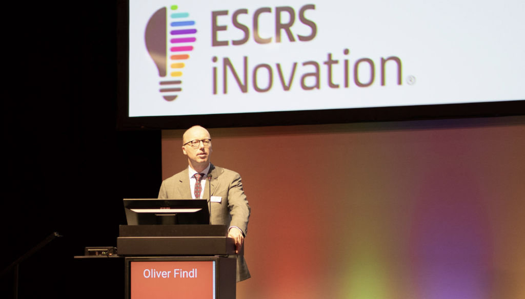
When Oliver Findl, President of the European Society of Cataract and Refractive Surgery (ESCRS), steps down at the end of this year, the Society shall remember his tenure as a time of both challenges and achievements—and, most certainly, will look back on the Viennese ophthalmologist’s two years at the helm with gratitude.
“It will be just like before the pandemic,” Oliver Findl mused a couple of weeks ago. He expected the ESCRS Congress in his hometown to be on par with the meetings before 2020—an understatement to say the least. This Congress puts all its predecessors in the shade.
Not only did ESCRS 2023 see record numbers of attendees, but its programme featured comprehensive insight into tomorrow’s ophthalmology, such as the iNovation Day. This is only the second year for the hugely successful initiative in bringing ophthalmic surgeons and industry together to highlight and debate the most recent innovations in ophthalmic surgery. And the Vienna delegates had a social blast that can hardly be topped: a Grand Ball held in the Hofburg, the palace where the Habsburgs ruled over a vast empire. This and much more was Oliver Findl’s doing.
Professor Findl, Chief of the Department of Ophthalmology at Hanusch Hospital in Vienna, is one of Europe’s most experienced cataract surgeons with more than 18,000 surgeries under his belt (not to speak of his retinal and corneal interventions). He completed post-doctoral research at Harvard University and is a constant on The Ophthalmologist’s “Power List”.
He has left his mark on the ESCRS in several fields. Regarding ongoing education—one of the Society’s primary aims—he has contributed not only as co-chair of the Clinical Guidelines Development Group for cataract surgery but has also encouraged many other initiatives, such as setting up a common e-learning platform to improve access to educational material.
Oliver Findl supported the creation of a new Digital Health Award that recently made its first grant. He helped establish an online IOL calculator that now includes toric lenses. Under his guidance, the ESCRS purchased a state-of-the-art surgical simulator from Haag Streit that has already supported skills development to scores of trainees in Romania, Poland, and now, Austria (for Ukrainian trainees). The uptake and feedback are beyond expectations, and that splendid machine’s next stop on its “Tour of Europe” is the combined ESCRS Winter Meeting/Annual Congress of the German Society of Cataract and Refractive Surgery (DGII) in February 2024 in Frankfurt.
Under his leadership, the ESCRS has become a force for environmental and social conscience that is beyond compare in the world of ophthalmology, as sustainability and fighting climate change have been a driving force for the Austrian ophthalmologist for many years. He was instrumental in making the ESCRS Congress carbon neutral and in promoting sustainability as a central theme internationally. Prof Findl played a vital role in establishing EyeSustain with ASCRS and AAO, where key issues such as reducing OR waste can be investigated. He has also established the Sustainability Index for Disposables in Cataract Surgery (SIDICS) project—a tool developed to assist users in evaluating the sustainability of customised cataract packs used in medical facilities and provides insights into the environmental impact of different product configurations.
Current events have kept him moving—rapidly and effectively responding according to circumstance. For example, Oliver Findl led the ESCRS in supporting Ukraine, encouraging industry partners to donate crucial surgical supplies. In addition to allocating a six-figure sum from its own reserves, ESCRS reached out to fellow societies to donate funds. He led the initiative to provide free congress registration for Ukrainian surgeons and emphasised the importance of relieving hardship in developing countries. Under his leadership, ESCRS funding for projects in developing countries has increased fivefold. In addition to major projects in Mozambique, Malawi, and Jerusalem, ESCRS has supported smaller cataract projects in South Sudan, Nepal, and Bangladesh.
Since stagnation means regression—as the saying goes—there will be several tasks for Prof Findl’s successor and the ESCRS council to pursue and complete. Among other things, the Society plans to provide patients (the “lay public”) with a website on cataract surgery. And the heritage website is under construction—things to accomplish after a presidential term that will surely be remembered for decades to come.
H Burkhard Dick, Secretary of the ESCRS and chair of the department of ophthalmology at Bochum University, Germany.
Howard Larkin reports
Emerging digital technology holds the potential to improve surgical outcomes, increase workflow efficiency, train more surgeons to meet exploding global demand, and eventually even lower costs. But the substantial upfront cost of developing and deploying digital technology—and who will pay it—remains a significant barrier, according to panellists at a session on the future of the digital operating room at the second ESCRS iNovation Day.
“Digital visualisation will be a really important factor. It’s not only about ergonomics for the surgeon: it’s about teaching, it’s about good documentation, and it’s especially about getting a lot of information into the visual field of the surgeon,” said Professor Oliver Findl in an introductory video. The session examined several aspects of what will be needed to fulfil the potential of the digital OR.
Impact of the digital OR
Streaming digital technologies that identify patients using biometric data gained in preoperative tests, project guides for toric lens alignment and other purposes, and live OCT and other imaging data are already available intraoperatively, noted session co-chair Professor Burkhard Dick. But uptake is limited, with only about 15% of surgeons using digital intraoperative marking systems. Infrastructure that supports data access and integration—and trust in technology to improve patient care—will be key for reaping the potential benefits in the short term, panellists said.
“Data is becoming more and more important in the OR, interconnectivity of systems is becoming more important,” said Zeiss’s Frank Seizinger. “The systems are talking to each other; a biometry system is talking to a microscope, is talking to a phaco machine, and giving the surgeon all the information on demand as they need it.” Over the next 10 years, integrating this type of clinical data will drive better outcomes and efficiency in the clinic. Adding digital patient questionnaires can shed light on patient needs and medical histories help improve decision-making. In addition to improving patient outcomes and satisfaction, such information could help increase workflow efficiency and throughput, he noted.
Alcon’s Seba Leoni added that reducing variability is paramount in cataract surgery, particularly with premium lenses. “The ability to have highly reliable diagnostic devices that ultimately enable seamless computation of the data and transmission into the operating room in a visual manner that allows for image guidance can really help improve surgeon precision, reduce variability, and deliver that [desirable] surgical outcome.” He cited research showing improved outcomes for surgeons who use digital markers rather than ink markers for aligning toric lenses, with a difference of about three degrees of alignment. Digital markers also eliminate the extra step of manually marking the astigmatism axis, increasing efficiency, Leoni added.
“Digital marking is a no-brainer that improves efficiency and outcomes,” said session co-chair Dr David Chang. So why isn’t it used more?
Among the challenges have been connectivity issues, Leoni said. A few years ago, when digital markers first appeared, data were mostly transferred by thumb drive. Then local area networks took over, and now the digital cloud. “We are moving in the right direction,” Leoni said. Uptake of existing technology roughly correlates with market penetration of premium lenses, suggesting that those with the biggest stake in increased precision are most likely to use it.
Dr Roger Zaldivar sees integrating digital data as essential to keep up with growing demand for services. He sees simplifying processes for not only doctors but also their staff as the key to improving efficiency and outcomes.
Dr Chang noted that most digital technology is predictive, leading to better outcomes through better preparation and execution. He cited the Light Adjustable Lens that can digitally change refraction after surgery as an extension of the digital OR that improves outcomes. “It’s a little daunting for those of us doing this for so long to think that someone a month out of training can get a fantastic result. … It’s a nice complement to all the digital technology we have in the OR.”
Digital visualisation
Improved and augmented visualisation is among the most important potential benefits of the digital OR, said Dr Damien Gatinel. Delicate procedures in the anterior chamber, such as placing endothelial grafts, as well as glaucoma and vitreoretinal procedures, would all benefit from enhanced stereoscopic view. Beyond this, augmented reality features, such as alarms when an instrument gets too close to fragile tissues, could enhance surgical precision and outcomes.
Prof Dick, who has used several advanced visualisation systems, agreed they are extremely valuable for delicate procedures. But cost is still an issue, especially for broader use.
“The systems are beautiful, but they are really expensive. If you are doing cataract surgery, how do you add enough value? What will it take to bring the cost down?” Dr Chang asked.
Improved ergonomics and better visualisation are advantages that once surgeons experience, they do not want to go back, industry panellists said. In addition, the cost of electronics tends to drop over time, which should make visualisation and other digital technologies more affordable as the technology develops, said Heidelberg’s Benedikt Wurm.
AI and robotics
The prospect of designing and programming a robot that can do everything surgeons can do is daunting, Dr Chang said. He likened the challenge to teaching a helicopter to fly on its own. No one could program it to anticipate every challenge, but teaching the helicopter how to correct problems as they occurred could make the project successful. “[With] machine learning, perhaps combined with a robotic system, you actually could succeed—that’s what intrigues me about it.”
Dr Zaldivar suggested taking smaller steps toward a robotic future may be the way forward. “For example, why do some surgeons have to retire after a certain [number] of years when they can still control and monitor a robot and still do surgery? There are things we can do heading in that direction.”
Dr Gatinel distinguished between autonomous and controlled robotic surgery, noting that while robots can be trained to do very precise procedures such as a capsulorhexis, paradoxically, placing a speculum is more difficult for a machine but easier for a human because the patient is anxious and may move their head in the process. “Removing the human completely? I don’t think is what we want or what patients want.”
Additionally, more and better data from digitally enabled diagnostic and surgical instruments will be needed to support machine learning, Wurm noted. “Digitalisation of the OR gives us an enabling technology to then develop semi-autonomous or even autonomous technologies.” Standardising such data is equally important, Dr Zaldivar said.
Training and education
AI and robotics also have a role in coaching and training surgeons, Leoni said. Using remote learning, Alcon’s phaco development programme has helped train 4,000 surgeons who have delivered more than 8 million procedures worldwide, he said. More recently, the company is developing a virtual training simulator in which students around the world can join a virtual operating room and look through the microscope as an instructor explains what they are doing in the procedure.
Cost, though, is an issue. However, much technology exists that can facilitate distance learning, including Zoom and inexpensive cameras.
Even so, 60% of participants polled for the session identified cost as the major barrier to implementing digital technology. A combination of reduced costs and proven benefit will be needed to overcome resistance, panellists said.
Removing unused tools from surgery trays could cut costs and waste.
Howard Larkin reports
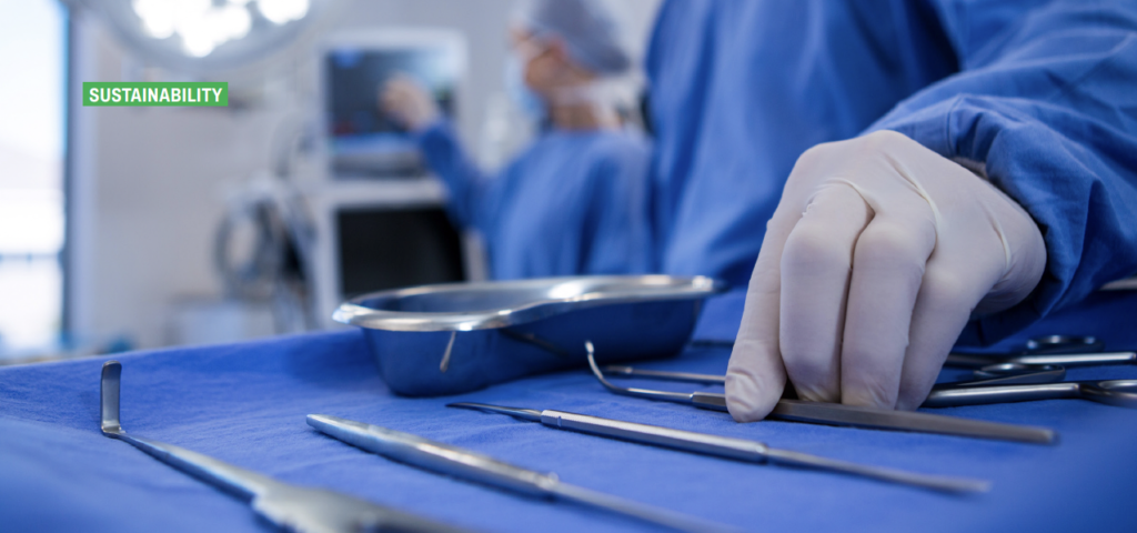
Identifying and removing unused surgical tools from high-volume surgical trays could save money and reduce the environmental impact of ophthalmic surgery, said Dr Victoria Liu of the University of Ottawa Eye Institute, Canada. The savings would mostly come from reducing processing and sterilising costs, as well as those for replacing damaged surgical instruments.
In a formal audit of 85 cataract surgeries by multiple surgeons at the institute, Dr Liu found about half of the tools in its cataract surgery trays were not used in a typical surgery. Overall, surgeons used a median of 46% of the tools per surgery, ranging from 32% to 59%. Seven of 22 tools on the tray were used in less than 2% of surgeries, with three going entirely unused.
Dr Liu estimated the total cost of these instruments, which did not include phacoemulsification tools, at more than $6,800 Canadian. Removing the eight tools used in less than 10% of surgeries would cut that total by about $2,470. Over just 50 trays, savings on the upfront cost of instruments would exceed $120,000. Actual savings would accrue through reduced handling, sterilisation, and replacement costs. Cataract surgery volume at her centre is about 8,500 cases per year.
An audit of corneal surgery trays for Descemet stripping automated endothelial keratoplasty (DSAEK) produced similar results, Dr Liu reported. A mean of just 32% of 32 tools on the tray was used per surgery, ranging from 22% to 38%, with 41%—or 13 instruments—used in no surgeries at all. The total cost of these 32 instruments is more than $12,000, so eliminating the unused tools for the more than 110 annual DSAEK procedures could save significant money.
However, Dr Liu acknowledged the study samples were small, adding result confirmation and the impact in the OR must be evaluated before making any changes to surgical trays. The audit project also demonstrated the complexity of opinions among multiple shareholders, including surgeons, nurses, and clinical care leaders, and generated resistance to making changes. “We are pending further feedback prior to making corneal tray changes.
Also, these costs are predicted, and the actual impact will need to be calculated after any future changes, Dr Liu added. However, while the environmental outcome is difficult to assess quantitatively, the impact on OR activities and financial waste can be calculated.
“Awareness and intervention to decrease unused surgical tools are important to reduce environmental costs in the OR, especially in the context of climate emergency and sustainability,” Dr Liu concluded, noting optimising surgical trays may also have a positive impact on OR procedures, patient care, and patient safety.
Dermot McGrath reports
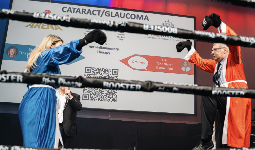
When a jackal is confronted with a jackhammer, the only possible outcome is a violent struggle for dominance.
And while there was thankfully no blood left on the canvas, the audience at the Arena debate on microinvasive glaucoma surgery (MIGS) versus selective laser trabeculoplasty (SLT) were still treated to a fiery contest as Karl “The Jackal” Mercieca from Germany slugged it out against Carlos “Jackhammer” Traverso from Italy.
Making the case for MIGS, Karl Mercieca came hurtling out of the blue corner with a volley of persuasive arguments.
“MIGS is way better than SLT. It can be combined with phacoemulsification, and the IOP lowering potential is way better than SLT,” he said. “If you look at the long-term results, with one procedure, you can get consistent pressures over a much longer period. With SLT, you need to repeat and repeat and repeat, and not all patients respond.”
Barely pausing for breath, Dr Mercieca followed up with another devastating combination.
“People say that SLT is cost effective and non-invasive, and so forth. But actually, it’s not that cost effective at all. If you look at MIGS combined with phaco, for example, the quality of life is better for your patient, and it’s actually more cost effective.”
The real appeal of MIGS is its versatility, argued Dr Mercieca.
“MIGS doesn’t mean one procedure—you have got so many options to choose from. The other thing is it doesn’t depend on having a nice open angle, whereas SLT can only be used in eyes with large, wide angles. MIGS can be used in other situations, especially combined with phaco, whereas SLT is not indicated in uveitis, narrower angles, and so forth. But the true beauty of MIGS is you can really tailor it. It’s truly personalised medicine for your glaucoma patients,” he concluded.
The Jackhammer fights back
In the red corner, the “Jackhammer” Traverso took the blows without complaint and decided to come out all guns blazing.
“First of all, what is the spelling for MIGS? If you are using MIGS, this means ‘my income grows steadily,’” he said to laughter from the assembled audience.
After this opening uppercut, Dr Traverso continued his verbal assault.
“Let us face it—MIGS are minimally effective. There is no comparable risk when we look at SLT,” he said. “With MIGS, we have seen endophthalmitis, choroidal issues, [etc.]. SLT is also doable without using the gonioscope. SLT is really not expensive, it is somewhat repeatable, and as a primary treatment, it is not only effective, it’s ethical. It is evident that there is no evidence whatsoever that trabecular MIGS are really working.”
Dr Mercieca responded that Dr Traverso’s arguments were strictly in the featherweight category.
“You are in the wrong boxing category because SLT is comparable to drops,” he said. “We are here at ESCRS with cataract surgeons who want to combine something with a cataract removal to get effective results, not something which they have to wait six weeks to see something positive.”
Dr Traverso was having none of it, delivering another stinging financial argument to his opponent: “Yes, of course, combine MIGS with phaco, so you get two surgical fees in 30 seconds. Who is going to pay for this?” he asked.
Dr Mercieca was quick to respond, adding long-term data showed that MIGS is a cost-effective procedure. “I would like to quote one Carlos Traverso back in 2014 who said, ‘MIGS is definitely the way forward and an alternative to current treatment with really expanding horizons’,” he said.
However, Dr Traverso wasn’t taking this potential hammer blow lying down.
“Yes, but those were the early data. We were part of the very first trial on this trabecular stent, and we participated enthusiastically. But what are the long-term results published in the literature? If it’s not confirmed by data, what is that for? My income grows steadily if I use MIGS, but I’m not there for income. I’m there for the patient’s well-being. So, I think that as a first step, SLT is really the only way out,” he said.
After some more lively trading of verbal blows in a hugely entertaining contest, both contestants retreated bloodied but unbowed to their corners and awaited the verdict of the audience present.
Summing up, the referee, Leon “The Lion” Au thanked both opponents for a lively debate and announced Dr Mercieca as the winner on points in a tight contest.
Dermot McGrath reports

National and international corneal transplant registries such as the European Cornea and Cell Transplantation Registry (ECCTR) are extremely valuable resources in determining the real-world success rates of different keratoplasty techniques and enabling benchmarking to drive quality improvement and reduce healthcare costs, among other benefits, according to Professor Mor M Dickman.
“Measuring is the basis for improvement: If we don’t know where we are in terms of our results and we have no basis for comparison with others, how shall we ever improve?” he said. “Registries such as ECCTR provide us with an opportunity to benchmark outcomes against colleagues in our clinic, country, and even across Europe. They provide a safe environment to learn from peers and encourage and incentivize improvement to benefit doctors and patients.”
Established in 2016, the ECCTR has now collected data on more than 20,000 transplants from 15 European countries, including information on the recipient, donor, and eye bank processing; transplant procedure; and two-year follow-up, including graft survival and failure and patient-reported outcome measures (PROMs).
Registry data such as that contained in the ECCTR constitutes a mine of valuable information that benefits practitioners and patients, Prof Dickman said.
Some key findings from the ECCTR include a mean recipient age for a corneal graft of 70 years, with Fuchs’ endothelial dystrophy as the primary reason for corneal transplantation, followed by graft failure, pseudophakic bullous keratopathy, and keratoconus.
“We can see that patient age changes according to diagnosis, with keratoconus patients considerably younger than Fuchs’ patients,” he said. “Patients with pseudophakic corneal oedema tend to be the oldest patients in the registry.”
Although, historically, Descemet’s stripping automated endothelial keratoplasty (DSAEK) was the dominant transplant procedure—followed by penetrating keratoplasty and Descemet membrane endothelial keratoplasty (DMEK)—the landscape has changed in recent years with DMEK now the dominant procedure.
“The adoption of deep anterior lamellar keratoplasty (DALK) remains limited, primarily due to increased cross-linking for keratoconus and the lack of specialised expertise in many centres,” Prof Dickman said.
The registry also highlighted that the primary objective of corneal transplantation is to improve vision, although other factors such as pain reduction, globe integrity, and infection debulking remain important objectives.
While the overall two-year transplant survival in the registry is 89%, Prof Dickman said survival is dominated by recipient diagnosis.
“Grafts performed for Fuchs’ dystrophy have better survival rates than grafts performed for bullous keratopathy,” he said. “Repeated grafts have a relatively poor prognosis, unfortunately.” Other poor prognostic factors are neovascularisation and a history of rejection.
The past decades have seen a profound shift to endothelial keratoplasty (EK). Nevertheless, the real-world data emerging from the registry indicates survival rates may not be as impressive as initially thought.
“What we have learned is that endothelial keratoplasty outcomes vary a lot. Looking at data from the Netherlands, we found a one-year graft survival of only 85% after DMEK compared with 95% for DSAEK. Almost all failures occurred in the first few months after surgery,” he said. “Although graft survival improved over time, for novice surgeons, a quarter of cases failed in the first few months after surgery.”
Going forward, Prof Dickman outlined the potential of registry-based randomised trials to try to inform best clinical practice in terms of corneal transplantation. One such trial currently underway compares two steroid regimens—dexamethasone and fluorometholone—in IOP elevation and endothelial cell loss. The trial will also evaluate cost effectiveness, with indefinite follow-up ensured via the registry data and in line with evidence-based medicine guidelines.
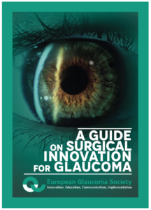 Glaucoma Day offered a full range of topics, from innovations in glaucoma surgery to how to handle post-phaco complications. The European Glaucoma Society used the occasion to debut a new book, A Guide on Surgical Innovation for Glaucoma, giving all attendees a free copy.
Two years in the making, the book is the result of systematic work by a panel of European glaucoma specialists led by three editors—Dr Luís Abegão Pinto (Lisbon), Dr Ingeborg Stalmans (Leuven), and Dr Gordana Sunaric Mégevand (Geneva).
The book is a response to the many innovations in glaucoma surgery in recent years. It allows readers to familiarize themselves with current procedures—both the minimally invasive glaucoma surgeries (MIGS) and bleb-forming devices—providing detailed descriptions of each, a summary of existing literature, and information on complications and caveats. In addition to telling readers what is out there, it addresses a need to create benchmarks for clinical trials to provide better data for comparing the risks and benefits of traditional and novel surgical interventions.
“The Guide offers an overview of the existing minimally invasive glaucoma surgery techniques but also aims to provide the reader with the tools for interpreting surgical studies,” said Dr Pinto, EGS Secretary. “Finally, or should I say, critically, it also aims to provide the standard for research on the field, hopefully raising the bar for future clinical trials.”
The editors gathered more than 250 questions from 50 European glaucoma specialists in 18 countries to name the top 10 clinical questions facing glaucoma surgery, which then went under systematic review. Where sufficient high-quality data was available, the editors included it but provided consensus statements based on clinical experience when sufficient data could not be found.
“We are not telling people what to do. We want to provide doctors with the data and the tools to make their own decisions,” Dr Pinto explained. “This book is intended to offer guidance for interventions popular enough to be widely used while avoiding bias towards techniques that are currently niche techniques.”
An important goal of the book is to create a framework of common standards for surgical trial design and reporting, making it easier to compare and judge future clinical studies using established benchmarks, he added.
The book will soon be available on the EGS website, www.eugs.org.
Dermot McGrath reports
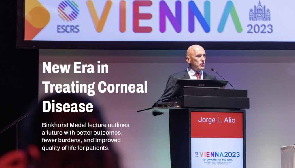
The treatment of corneal diseases is undergoing a paradigm shift, which will overcome many of the shortcomings of current approaches to dealing with corneal dystrophies and other potentially blinding conditions, said Professor Jorge L Alió in his Binkhorst Medal Lecture at the 2023 ESCRS Congress in Vienna.
“We are at the forefront of an exciting revolution in the treatment of corneal blindness, moving from the old paradigm of cornea tissue substitution to one where cornea tissue regeneration becomes the new standard,” Prof Alió said.
The need for new treatments is pressing, he stressed, noting that corneal blindness is the second most common cause of blindness worldwide, with treatment limited by the number of donors for corneal transplants.
“The shortage of corneas results in over 40,000 visually impaired people waiting for corneal transplants every year in Europe alone, with 10 million untreated corneal blindness patients globally and 1.5 million new cases of corneal blindness annually,” he said.
This number is rising due to the ageing population and corneal graft failure, which often occurs due to rejection, becoming even more likely with each successive graft and new indication.
“There is a need for new solutions to avoid tissue substitution, preferably immunologically neutral ones, independent from human donor tissue, with unlimited accessibility that will probably be more cost effective than corneal grafts in the long term,” he said.
Advanced stem cell therapies may be the game-changer everyone has been waiting for, added Prof Alió, who outlined some of the latest research in regenerative techniques for a wide range of corneal diseases.
Adipose-derived stem cells (ADSCs), for instance, have been shown to provide a viable cell source for stromal regeneration and repopulation in diseased corneas. ADSCs are typically isolated from adipose (fat) tissue obtained from the patient’s body, which reduces the risk of rejection or immune-related complications.
ADSCs can be induced to differentiate into epithelial cells or stromal cells in the laboratory and can help repair the corneal epithelium, stroma, or endothelium, depending on the specific corneal injury or disease.
These cells have demonstrated anti-inflammatory and immunomodulatory properties, which can be beneficial for reducing inflammation and immune responses in the damaged cornea. They may also stimulate endogenous repair mechanisms in the cornea by releasing growth factors and cytokines that promote tissue healing.
The first-in-human trials successfully tested the technique using ADSCs implanted into corneal stroma in five patients with advanced keratoconus.
Other approaches using mesenchymal stem cells (MSCs) have shown promise for treating ocular surface diseases such as dry eye, corneal burns, ulcers, and limbal stem cell deficiency (LSCD).
As well as epithelial repair, MSCs can be differentiated into corneal stromal cells in vitro and then transplanted into the corneal stroma to promote tissue regeneration. The cells can also be incorporated into tissue-engineered constructs or biodegradable scaffolds for corneal transplantation in thin corneas or cases where injection alone is insufficient to promote corneal regeneration, said Prof Alió.
“MSCs have a present and future critical role in the management of corneal epithelial failure due to limbal stem cell deficiency. Severe forms of dry eye disease have already been improved with MSC therapy in open-label clinical trials, with randomised controlled clinical trials to follow,” he said. “Cell-free therapy such as PRP—especially with MSC-extracellular vesicles (such as exosomes)—could be a future better option than MSCs, provided the many existing challenges can be solved.”
Looking to the future, Prof Alió predicted corneal organoids could potentially solve the corneal graft tissue shortages.
He explained the corneal organoids are bio-constructs made with corneal epithelium tissue, stromal laminas from non-human origin, and endothelial tissue shaped with 5D printing techniques to deliver customised corneal optical power.
“This will improve patient quality of life and reduce the economic burden of corneal transplants on healthcare,” he said. “Furthermore, we expect these bio-constructs to provide better therapeutic outcomes over donor corneas, such as avoiding allograft rejection.”
Dermot McGrath reports
Ukrainian ophthalmologists continue to push their medical and surgical skills to the limit in dealing with the devastating impact of eye injuries incurred among both the civilian population and military personnel in the ongoing war with Russia.
The horrific extent of those injuries—and the various strategies employed by Ukrainian ophthalmologists to deal with them—were brought forcibly home to delegates during a special session on ocular trauma at the 2023 ESCRS Congress in Vienna.
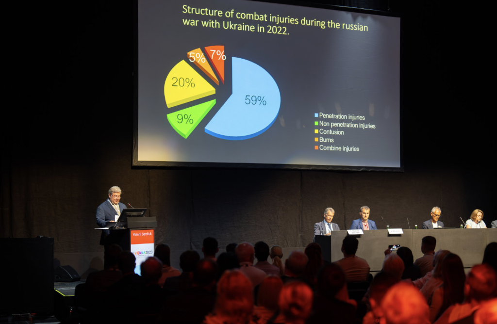
Eye trauma is estimated to account for up to 13% of all injuries in modern warfare, and the war in Ukraine is no exception, said Dr Valerii Serdiuk, who recounted his experience of combat surgery over the past nine years.
“Evolution in warfare tactics means that anti-personnel mines and various explosive devices—both improvised and produced by industry—have become the main causes of eye combat trauma in all military conflicts,” he said. “Other causes include wounds from firearms and accidents.”
Thousands of military and civilian patients have been treated by the eye specialists at Dnipropetrovsk Hospital since the outbreak of conflict, said Dr Serdiuk, adding his teams typically encounter a high level of complex ocular polytrauma, often in association with other head, neck, face, or systemic injuries.
The advanced fragmentation weapons used in modern conflicts results in a high rate of ocular trauma, with binocular injuries in 34% and penetrating injuries in 42% of cases.
“The high percentage of eye injuries in the first months of the war was related to the shortage of protective eyewear and a lack of awareness on the part of military personnel about the threat to their visual health,” he said.
In this regard, he noted eight eviscerations/enucleations were performed from 2014 to 2021, but seven of those were carried out before September 2014, when protective eyewear use became widespread.
Dealing with ocular polytrauma requires a clear strategy on the part of the surgeon, Dr Serdiuk said. In penetrating and blunt eye injuries, there is usually combined damage to the anterior and posterior segments, including the cornea, iris, and retina, causing significant and diverse clinical and functional disorders in the injured eye.
Rapid intervention is also important to improve the prospects of saving sight in patients with severe ocular polytrauma, Dr Serdiuk added.
The complex nature of the ocular injuries facing surgeons in Ukraine was also described in detail by Professor Nadiia Ulianova, who reported on her experience treating combat victims at the Filatov Institute of Eye Diseases and Tissue Therapy in Odesa.
“Modern combat trauma is particularly severe and requires complex reconstructive treatment,” she said. “The optimal timing for pars plana vitrectomy (PPV) for open globe injury varies from one to four weeks from the moment of injury and eight weeks post-injury for keratoplasty.”
Prof Ulianova outlined the challenges of performing vitrectomy in traumatic injury cases.
“The most common indications for vitrectomy in cases of ocular trauma are vitreal haemorrhage, retinal detachment, intraocular foreign bodies, and macular holes,” she said. “All of these indications are usually present in severe combat injuries.”
Although vitrectomy in trauma cases should ideally be performed as soon as possible, the reality is logistical difficulties in displacing wounded individuals from the frontlines and ensuring treatment of other life-threatening injuries often affect the timing of ocular surgery, she added.
Prof Ulianova listed some specific features of eye trauma due to modern combat, including extensive open globe injuries and multiple foreign bodies in the cornea. She highlighted strategies to deal with some of these scenarios, including using keratoprostheses, amniotic membrane, and soft contact lenses—either individually or in combination with PPV to try to rehabilitate the ocular structures and save vision.
The winner of this year’s John Henahan Writing Prize was announced during the opening ceremony of the Congress. Congratulations to Siyin Liu MD.
Here is the prize-winning essay.
By Siyin Liu MD

Medicine is at a critical inflection point for artificial intelligence (AI). With a whopping 3,327 new AI companies in the mix and a projected $37 billion splurge on AI by 2025, this tech is drastically transforming every industry, including healthcare.
Ophthalmology, with its rich imaging data, presents an ideal setting for training algorithms in image recognition, segmentation, and disease detection. Current focus lies on prevalent ophthalmic conditions like diabetic retinopathy (DR), age-related macular degeneration (AMD), and glaucoma, leveraging large, standardised imaging data sets. The COVID pandemic accelerated the integration of AI into tele-ophthalmology, exemplified by the FDA-approved autonomous diagnostic device for DR, enabling point-of-care diagnosis without human oversight. Challenges exist in AI research for anterior segment diseases due to non-uniform slit-lamp images and limited data sets. Nevertheless, recent advancements demonstrate AI’s potential in the anterior segment, including early detection of keratoconus, post-refractive surgery ectasia screening, and diagnosis of infectious keratitis.
AI in ophthalmology has primarily focused on image-based deep learning. Yet, the adoption of electronic health records has unveiled the untapped potential of unstructured free-text data. Natural Language Processing (NLP) and algorithmic rule-based text extraction techniques have shown promise in leveraging this data to enhance care delivery and for big data analysis, predictive modelling, cohort identification, and stratification. NLP also standardises specialised ophthalmic terminology, facilitating interactions among healthcare providers and patients. With predictions that 85% of customer interactions will be managed without human agents by 2025, NLP-based chatbots hold tremendous potential in triaging symptoms, monitoring treatment adherence, and providing support in areas with limited ophthalmology services.
AI chasm
A core challenge in applying AI is the clinical validation of recently developed concepts and tools. Clinical AI research faces limitations due to retrospective design, leading to biased algorithms “overfitted” to specific data sets. The conventional approach of pitting AI against clinicians may not demonstrate real-world performance, as its realistic application likely involves interaction between clinicians and algorithms. Clinicians, the end users, must grasp AI’s strengths and limitations to foster mutual learning. Envisioning AI’s role in high-risk real-time situations like surgery, where surgeons adapt their approach on the fly, is challenging.
AI regulatory approval is another challenging obstacle. Many algorithms rely on complex and opaque mathematical models, often referred to as “black boxes.” The lack of transparency raises concerns about data mishandling and understanding of the algorithms’ inner workings. Agencies like the FDA/EMA require extensive transparency in scientific methods, but researchers and companies may hesitate to expose proprietary algorithms publicly due to potential financial risks. Further, without a clear understanding of algorithmic processes, AI may struggle to gain patients’ trust and approval. Would it be worse for patients to be misdiagnosed by a human or a machine? What if the algorithm had demonstrated superior performance in research settings? Ultimately, trust and confidence in algorithmic decision-making play a pivotal role.
Overall, more rigorous work is needed to combat the disparity between AI hype and application in healthcare, the so-called “AI chasm.”
The glass cage
The integration of AI into healthcare runs the risk of stripping medicine of its human touch. With advancing algorithms, we may see clinics where machines take the lead, like a macular clinic where an OCT machine decides on intravitreal anti-VEGF injections for AMD without ophthalmologist involvement. Even with fancy AI voice synthesizers like Siri, the empathy and intuition of the doctor-patient relationship diminishes. After all, to these machines, patients are reduced to mere data points where efficiency and cost-effectiveness are the sole measures of success. Further, overreliance on AI-powered automated diagnosis or treatment decisions may stunt the clinicians’ development of critical thinking and decision-making skills, which may, with time, add them to the list of skills lost due to technology.
Biases embedded in the training data can perpetuate inequalities by providing inaccurate or inadequate recommendations for certain groups or populations. With training data predominantly derived in the Western world, algorithms may struggle to effectively generalise to diverse groups. Unchecked, AI can reinforce and amplify existing disparities in healthcare outcomes. Also, the ethical implications of data ownership and privacy protection arise in an era of round-the-clock data collection from gadgets and wearables. Algorithms may exploit this data and stigmatise the chronically ill or those who don’t fit the “healthy lifestyle” mould, potentially leading to unjust financial and health penalties, such as reduced access to insurance.
The rise of AI
We are told AI-powered healthcare is not about replacing ophthalmologists, but rather augmenting their knowledge and expertise. The integration of genomic data, lifestyle factors, and imaging will lead to AI-powered decision support systems that can guide targeted therapies and tailored interventions. Although AI’s current capabilities fall short of the hype, overcoming challenges could unlock its potential in achieving the holy grail of personalised medicine. Yet, the question remains whether the rise of AI lead to the “machinification” of medicine.
Dr Liu is in Specialty training year 4 at UCL Institute of Ophthalmology, London, UK.
Email: siyin.liu@ucl.ac.uk